Комментарии:

THANK YOU MAN YOU REALLY HELPED ME
Ответить
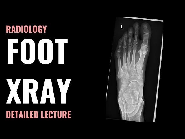
I have just had an XRay for my great toe - PIP joint due to a small bump and mild pain. I had a quick look at the Xray after it was done and it looked like there was significant subchondral sclerosis, but from the lateral view there was (from what i could see), no cartilage narrowing. What does this mean? I have 2 weeks to wait for the report and I'm dead anxious now.
Ответить
M Wishing everytime i see you feet part ofs..
Ответить
Unfortunately, most radiographers in from India include the lower 1/3rd of the leg in foot xrays, as shown in the series. This increases the radiation dose by 2 while reducing the image contrast through scatter. Scatter dose to male gonads is increased by about 4 when compared to accurately collimated images. The standard projection for the AP is using an up tilt of 15 degrees which is never done in India.
Ответить

![[청음 & 청음평가] Verity Audio Leonore 청음영상 (feat. GoldMund Telos 440, Mark Levinson No.534, Mimesis 37S) [청음 & 청음평가] Verity Audio Leonore 청음영상 (feat. GoldMund Telos 440, Mark Levinson No.534, Mimesis 37S)](https://rtube.cc/img/upload/RXc4bUJEUjg3SzU.jpg)
![Black Bean Instant Noodles Mukbang [Dorothy] Black Bean Instant Noodles Mukbang [Dorothy]](https://rtube.cc/img/upload/VW9wbzlUR1pNdFU.jpg)


